Conditions & Treatments

Benign skin tumours are growths caused by abnormal proliferation or collection of cells in the skin. These tumours are benign and they do not turn cancerous. Some common benign skin growths are moles, skin tags and seborrhoeic keratosis.

Note:  Leave the name of the tab "blank"if you do not wish to show this tab.
Melanocytic naevi (Moles)

​​Most moles occur in childhood, ado​lescence and early adulthood. They are due to abnormal collection of pigment producing cells known as melanocytes in the skin. Most moles are dark and pigmented but some moles may be lightly pigmented or may even be flesh coloured. Moles are small (< 5mm) flat and pigmented when they appear but the clinical appearance changes with the life cycle of the mole. With advancing age, they become raised and dome-shaped and often appear lighter. These changes do not signify cancerous changes and except for cosmetic concerns, do not require removal. Occasionally, changed moles may be a cause for concern if they develop irregular border, bleeding or sudden increase in size and you should consult a dermatologist.​

Treatment

Moles are often removed on cosmetic grounds. The choice of treatment is surgical excision. The mole may be sent for histological examination for confirmation of diagnosis or to rule out an atypical mole or cancer. After excision, there will be stitches in place which will be removed after 7 to 14 days, depending on the site of the mole. The mole will be replaced by a line scar.

Skin tags
Skin tags

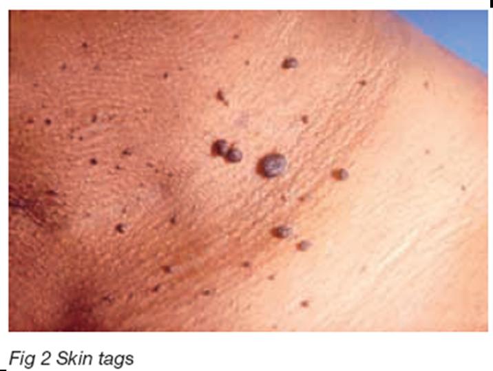
Skin tags (Fig 2) are harmless skin-coloured or brown growths, which commonly occur on the neck, underarms, groin, and eyelids. They vary in size fr​om less than 1mm to as large as 10mm. They are usually asymptomatic and often occur as multiple lesions.

Treatment

​Skin tags can be easily removed, if so desired, by snip excision and electrocautery. This is done under topical anaesthesia such as using EMLA cream. After the surgery, there will be superficial wounds which heal in 4 to 7 days. There may be some post inflammatory darkening after the wounds heal, but this will usually fade with time, over the next few weeks.

Seborrhoeic keratosis (seborrhoeic warts)

​​These lesions are superficial skin overgrowths which appear in increasing numbers with age. In early lesions, they are brown and slightly raised; however, they may become fairly large and pigmented nodules. They are commonly located on the face and can also be seen on the trunk, groin and sun-exposed areas. Sebo​rrhoeic keratosis (Fig 3) is benign lesion and do not transform to skin cancer.

Treatment

Cosmetic removal of seborrhoeic keratosis can be performed in various ways, depending on the clinical type. Early seborrhoeic keratosis can be treated with liquid nitrogen. When multiple, seborrhoiec keratosis is best treated with electrocautery.

Sebaceous hyperplasia

​​Sebaceous hyperplasias are yellowish lesions often seen on the cheeks. They have a characteristic central dimpling. This benign tumour is due to an increase in proliferation of oil glands just below the surface of the skin.

Treatment

Sebaceous hyperplasia may be treated with electrocautery or laser. These treatments will flatten the lesions; however there is a risk of scarring, post inflammatory darkening and recurrence following the treatment.

Syringoma
Syringoma

​​Syringomas (Fig 4) are benign tumours due to the abnormal proliferation of sweat ducts in the skin. They are flesh coloured papules, often located just below or around the eyes. A family history may be present.

Treatment

Syringomas may be treated with laser or electrocautery. The treatment will flatten the lesion but there is a risk of scarring and recurrence of the lesions following treatment.