Conditions & Treatments

​​​Chronic Venous Leg Ulcers _ Dr Mark Tang / Yr 2005

A venous leg ulcer located above the ankle
Fig 1 A venous leg ulcer located above the ankle.

Livideo vasculitis
Fig 2 Livideo vasculitis.

Ischaemic foot ulcers and Diabetic Mellitus
Fig 3 Ischaemic foot ulcers and Diabetic Mellitus.

What are venous leg ulcers? Venous leg ulcers (Figure 1) are the commonest cause of chronic, non-healing ulcers of the leg. They occur in both adult men and women and can take a long time to heal. There are many other causes of chronic leg ulcers (Figure 2, 3) and proper evaluation by a doctor is necessary to make the diagnosis.

Note:  Leave the name of the tab "blank"if you do not wish to show this tab.
What causes venous leg ulcers?

​​The blood supply of the leg consists of blood vessels called veins that return blood from the lower legs to the heart, assisted by valves that ensure that the “one-way” upward flow of blood. In many patients with venous leg ulcers, these valves are not working properly, causing blood to “pool” in the lower legs. This can lead to swelling and darkening of the skin, and eventually to venous leg ulcers (Figure 4).


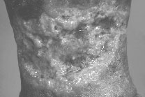
Fig 4 Venous Leg Ulcer - The skin of the leg can turn dark brown or red.
The ulcer is usually wet and weepy, with heavy discharge.

What are the risk factors for venous leg ulcers?

​​Some of the risk factors include patients with a past history of a blood clot in the leg veins (deep vein thrombosis), lower leg injury, varicose veins, obesity, smoking and prolonged standing. Patients who have difficulty moving or walking can also get venous leg ulcers because of loss of calf-muscle pump action which is important for venous blood circulation.

What are the signs and symptoms of venous leg ulcers?

​​Most patients with venous leg disease complain of aching and swelling of their legs, especially at the end of the day. The skin of the leg can turn dark brown or red, or there can be an itchy red rash with scaling and crusting (Figure 5). Small or large varicose veins may be seen (Figure 6).

Stasis eczema - itchy red rash with scaling and crusting on the skin
Fig 5 Stasis eczema - itchy red rash with
scaling and crusting on the skin.

Varicose veins shown on the lower leg
Fig 6 Varicose veins shown on the lower leg.

Venous leg ulcers are often located just above the ankle. They are usually wet and weepy, with heavy discharge. They can be painful and tender.

What is the treatment?

​Once the diagnosis is confirmed, venous leg ulcers can be effectively treated by good wound care and the use of compression therapy that can improve the venous flow and decrease the swelling of the legs. Compression therapy usually consists of special “elastic” bandages that must be applied by trained medical personnel. The arterial blood supply of the lower limbs must be adequate before compression therapy can be used.

What is the best way to prevent venous leg disease?

​​Once the leg ulcer is healed, it is important to prevent recurrence by wearing support stockings. It is important to replace the support stockings every 3 to 6 months or once the elasticity of the stockings are worn out. Other important measures include good skin care, regular exercise, weight management, stopping smoking and leg elevation above the level of the heart when lying down.​